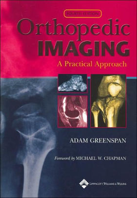
(PDF) Orthopedic Imaging – A Practical Approach 4th Edition by Adam Greenspan
$28.00
Download instantly Orthopedic Imaging – A Practical Approach 4th Edition by Adam Greenspan, Laura Pardi Duprey, Michael W. Chapman. It is ebook in PDF format.
ISBN-10: 0781750067 ISBN-13: 9780781750066
Preview
This is the PDF eBook version for Orthopedic Imaging – A Practical Approach 4th Edition by Adam Greenspan, Laura Pardi Duprey, Michael W. Chapman
Table of Contents
1: The Role of the Orthopedic Radiologist
2: Imaging Techniques in Orthopedics
3: Bone Formation and Growth
4: Radiologic Evaluation of Trauma
5: Upper Limb I: Shoulder Girdle
6: Upper Limb II: Elbow
7: Upper Limb III: Distal Forearm, Wrist, and Hand
8: Lower Limb I: Pelvic Girdle and Proximal Femur
9: Lower Limb II: Knee
10: Lower Limb III: Ankle and Foot
11: Spine
12: Radiologic Evaluation of the Arthritides
13: Degenerative Joint Disease
14: Inflammatory Arthritides
15: Miscellaneous Arthritides and Arthropathies
16: Radiologic Evaluation of Tumors and Tumor-Like Lesions
17: Benign Tumors and Tumor-Like Lesions I: Bone-Forming Lesions
18: Benign Tumors and Tumor-Like Lesions II: Lesions of Cartilaginous Origin
19: Benign Tumors and Tumor-Like Lesions III: Fibrous, Fibroosseous, and Fibrohistiocytic Lesions
20: Benign Tumors and Tumor-Like Lesions IV: Miscellaneous Lesions
21: Malignant Bone Tumors I: Osteosarcomas and Chondrosarcomas
22: Malignant Bone Tumors II: Miscellaneous Tumors
23: Tumors and Tumor-Like Lesions of the Joints
24: Radiologic Evaluation of Musculoskeletal Infections
25: Osteomyelitis, Infectious Arthritis, and Soft-Tissue Infections
26: Radiologic Evaluation of Metabolic and Endocrine Disorders
27: Osteoporosis, Rickets, and Osteomalacia
28: Hyperparathyroidism
29: Paget Disease
30: Miscellaneous Metabolic and Endocrine Disorders
31: Radiologic Evaluation of Skeletal Anomalies
32: Anomalies of the Upper and Lower Limbs
33: Scoliosis and Anomalies with General Affect on the Skeleton


